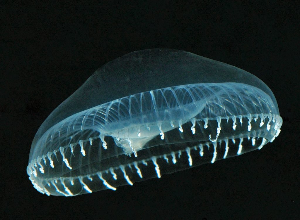
The bioluminescent jellyfish Aequorea victoria is the source of green fluorescent protein (GFP) Credit: Sierra Blakely, Wikimedia Commons
My research team, which is funded by the National Science Foundation (NSF), explores bioluminescence—the biological production of light by natural chemical reactions. Specifically we focus on the evolutionary origins of bioluminescence in Motyxia, the only millipede genus in California that is bioluminescent.
Interestingly, Motyxia are blind, and so their visual signaling can only be seen by members of other species, such as predators. In addition, Motyxia produce hydrogen cyanide, an extremely poisonous gas as a chemical defense. Some of our research indicates that Motyxia’s bioluminescence serves as a warning signal to deter nocturnal mammals from eating these highly poisonous millipedes.
By helping to reveal the evolutionary origins of bioluminescence, we can better understand and investigate how other complex traits arise in nature. In addition, bioluminescence research has a history of offering many proven and potential societal benefits in fields ranging from national defense to medicine. Some examples:
- Bioluminescence is also called “cold light” because the biochemical reaction that generates bioluminescent light is typically more than 90 percent efficient, meaning that only 10 percent of bioluminescent chemical energy is wasted as heat. By contrast, incandescent light bulbs are only 10 percent efficient! We could enhance the efficiency of our lighting systems by designing them to mimic natural bioluminescent reactions.
- The bioluminescent underbelly of the marine bobtail squid blends with background light from the water’s surface and so decreases the squid’s vulnerability to attack from predators dwelling below it. This natural camouflage offers the potential to inspire luminescent (glow in the dark) hulls for warships. The U.S. Navy is currently studying marine animals that use bioluminescence.
- All humans spontaneously release ultra-weak photon emissions and generate light through processes that are similar to bioluminescence in other animals. However, cancerous cells emit more light than do normal cells. This difference suggests that human luminescence may be used as a clinical tool to help diagnose illness and pinpoint the exact locations of cancerous cells.
- NSF-funded biologist Osamu Shimomura wanted to know what caused the jellyfish Aequorea victoria to glow green. One protein he found in the jellyfish, called green fluorescent protein (GFP), has revolutionized how scientists study cells. GFP is now widely used in biological and biomedical research as a fluorescent tag to help researchers track specific biological activities, such as the spread of cancer, the production of insulin and the movement of HIV proteins. And in 2008, Shimomura along with Drs. Martin Chalfie and Roger Tsien received the Nobel Prize in Chemistry for the discovery and development of GFP.
- The enzyme that is responsible for bioluminescence in beetles is used by researchers to carry out next-generation pyrosequencing—a fast, inexpensive method for sequencing genomes. In 2008, pyrosequencing was, for the first time, used to sequence the full genome of an individual human; the human was Dr. James Watson—the co-discoverer of DNA. Also, in 2008, pyrosequencing was used to sequence the complete genome of a Neanderthal.
- Enzymes responsible for bioluminescence are used by researchers to detect ATP (adenosine triphosphate), which is an essential substance for living cells.
- A version of a light-emitting compound from the seed shrimp Vargula (a small bioluminescent crustacean) is used by researchers to measure superoxide anions, which are a critically important component of metabolic systems that are difficult to detect in nature.
- Many types of physiological processes trigger changes in concentrations of intracellular calcium ions—an essential component of biological processes. One way to track intracellular calcium concentrations is to insert into cells a type of photoprotein known as aequorin, which is derived from the bioluminescent jellyfish A. victoria. Aequorin is highly sensitive and specific to calcium and—most importantly—emits light when it reacts and thereby signals calcium concentrations. In addition, it is non-toxic to most cells.
- Other photoproteins derived from diverse animals might be used someday to help understand other complex but fundamentally important biological dynamics. For example, Beroe, a comb jelly and Thalassicolla, a radiolarian, might also be used to help detect calcium ions, and Harmothoe, a scaleworm, might be used to detect iron ions.
Next time you’re up late and it’s dark outside (even better right after a summer rain), visit your local natural area—a moist gully or streamside are the best. Turn off your flashlight and allow your eyes become adjusted to the light. Blue ghosts, railroad-worms, luminous millipedes, and snail-eating firefly larvae are some of the bioluminescent organisms you might see. When you observe these fascinating organisms, consider how they emit light and why their ability to bioluminesce evolved. But importantly, just take a moment to quietly observe the nightlife and nature’s living light.
Continue reading →