millipede from Blacksburg, VA


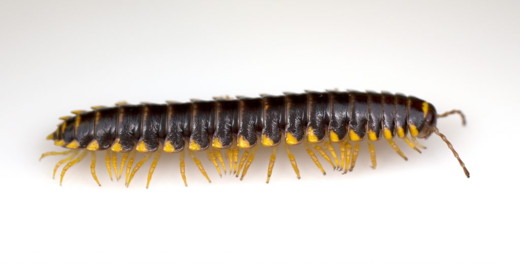
Appalachioria separanda calcaria (Blacksburg, VA)

Here’s a local cyanide-producing millipede from near Virginia Tech’s campus. In 1959, William Keeton (then a student at VT) described this species based on specimens   collected from Blacksburg, Riner and Radford, Virginia. This species is endemic to just a few spots in SW Virginia and S West Virginia, which are the only places in the world where this millipede occurs. (Thanks to Jamie Wahls, a graduate student in the Entomology Department, for sharing his discovery.)

Photo by P. Marek and T. McCoy (Canon 6D, 50 mm lens, 1/60 s, 5.6 f)

Keeton, W. T. (1959). A revision of the milliped genus Brachoria (Polydesmida: Xystodesmidae). Proceedings of the United States National Museum, 109: 1-58.

Posted in news | Tagged , , , , , | Leave a comment

Armored scale


Gloomy scale (Melanaspis tenebricosa), an armored scale in the family Diaspididae

Eric Day, Insect ID Lab Manager @ Virginia Tech, kindly shared these wonderful gloomy scales with us one day during Insect Biology Lab. This picture shows two female scales (top-center) with their protective covers removed. The scale insects are the pink round things, and you can see their hardened pygidial (tail) region on their right. The scales in this stage really don’t have legs, eyes or antennae.


Gloomy scale pried from its feeding spot

In this image, the female scale is on its side (tail on the right), and you can see its very long thread-like mouthparts that are used like a straw to drink the juices of the tree.

Posted in news | Tagged , | 1 Comment

Tiny scelionid wasp

ScelionidEM21
Parasitic wasp in the family Scelionidae (Telenomus Gryon sp.) Thanks István!  😉

James Wilson, graduate student here in the Department of Entomology at Virginia Tech working in Tom Kuhar’s Vegetable Entomology Lab, recently visited our lab with some of his parasitic wasps. This is an image of an unidentified species of Telenomus Gryon reared from stink bug eggs that we captured with our new microphotography system. It’s a tiny little thing, about 1 mm long!
(Canon 6D, 65 mm lens, 4x, 1/125s, f5.6 – stack of 10 images)

Posted in news | Tagged , , , | 2 Comments

Cyanide gland dissections

millipede cyanide gland
Cyanide gland, expertly dissected from Pachydesmus crassicutis by Tim McCoy. The large balloon shaped reservoir on the left stores the stable precursor mandelonitrile, which is squeezed into the thickened, oval-shaped reaction chamber on the right. In the reaction chamber, mandelonitrile reacts with an enzyme to produce cyanide and benzaldehyde. The noxious cocktail is finally exuded from the “ozopore” opening to the environment (tubular projection shown on the right). This structure is located internally with the precursor reservoir facing mediad (towards the heart & nerve cord) and reaction chamber oriented laterad (towards the face of a marauding predator).

Reference: Eisner, T., H.E. Eisner, J.J. Hurst, F.C. Kafatos, J. Meinwald. 1963. Cyanogenic glandular apparatus of a millipede. Science, 139, 1218-1220.

Posted in news | Tagged , | Leave a comment